| Project Leader | Toshio ANDO Professor, School of Mathematics and Physics, Kanazawa University |
||||
| U R L | http://www.s.kanazawa-u.ac.jp/phys/biophys/index.htm | ||||
|
|||||
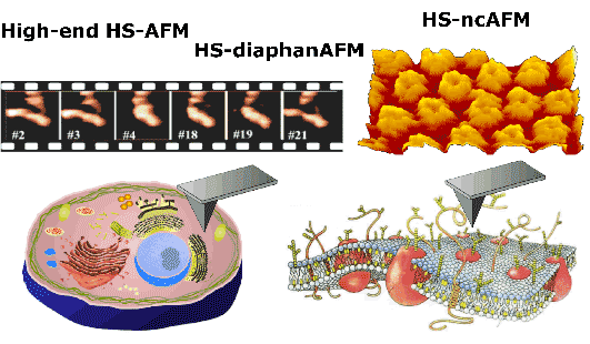
We have been leading the world in high-speed AFM. We have developed high-speed AFM which allows the direct visualization of the dynamic behavior of isolated biological macromolecules. However, even this most advanced instrument has not reached a level high enough for application to a vast range of samples. In this project, therefore, we aim at developing a high-performance high-speed AFM through improvements in the instrument and the development of new types of high-speed AFM which greatly expand the scope of target samples. Live cell membranes are extremely soft, so that they are significantly deformed through contact with the AFM probe tip, making it impossible to visualize molecular processes that take place on the membranes. To achieve this sort of visualization, we are developing high-speed noncontact AFM (or high-sensitive high-speed AFM) which prevents the deformation of live cell membranes. AFM is generally considered as microscopy for the observation of surfaces. However, it is possible to observe sub-surface structures in combination with ultrasonic techniques. Here, we introduce ultrasonic techniques to high-speed AFM to develop high-speed “diaphan-AFM” to observe the dynamic behavior of intracellular organelles. When cells are the target samples, the area to be observed is wide, requiring optical microscopic observations. Although designing a high-speed AFM apparatus that fulfills these requirements is not an easy task, we will overcome the difficulties to produce highly versatile high-speed AFM. Thus far, seventeen patents (both granted and pending) have resulted from the course of high-speed AFM development and have been licensed to domestic and oversea manufacturers. The development of prototype products is now in progress at the companies. We anticipate that “high-performance high-speed AFM”, “high-speed noncontact AFM” (or “high-sensitive high-speed AFM”) and “high-speed diaphan-AFM”, which we are attempting to develop through this project, will also go through several steps such as the production of prototypes by us, patent licensing, market research by manufacturers, and trial and evaluation by users. After going through these processes, final commercial products will be developed and eventually brought onto the market by several companies.

The Hokuriku Industrial Advancement Center